Perfect ARDMS Authentic SPI Exam Hub | Try Free Demo before Purchase
Wiki Article
What's more, part of that PracticeVCE SPI dumps now are free: https://drive.google.com/open?id=1V_UCURtI2-G4muVwdwMGYXL_h-sw8WnG
In modern society, everything is changing so fast with the development of technology. If you do no renew your knowledge and skills, you will be wiped out by others. Our SPI guide materials also keep up with the society. After all, new technology has been applied in many fields. So accordingly our SPI Exam Questions are also applied with the latest technologies to be up to date. You can free download the demos to check that how wonderful our SPI learning praparation is!
ARDMS SPI Exam copyright Topics:
| Topic | Details |
|---|---|
| Topic 1 |
|
| Topic 2 |
|
| Topic 3 |
|
| Topic 4 |
|
| Topic 5 |
|
SPI Materials, SPI Mock Exams
While SPI exam preparing for the Sonography Principles and Instrumentation (SPI) exam, candidates have to pay extra money when ARDMS introduces new changes. With PracticeVCE you can save money in this scenario as up to 365 days of free updates are available. You can also download a free demo to understand everything about PracticeVCE SPI Exam Material before buying.
ARDMS Sonography Principles and Instrumentation Sample Questions (Q92-Q97):
NEW QUESTION # 92
Which artifact is caused by defects in the crystals of the transducer?
- A. Ringdown
- B. Dropout
- C. Mirror image
- D. Side lobe
Answer: B
Explanation:
Comprehensive and Detailed Explanation From Exact Extract:
Defects in transducer crystals result in missing or weakened signals along the beam path produced by those elements, creating dropout. In array transducers, dropout typically appears as vertical or horizontal dark zones depending on which elements are affected.
According to sonography instrumentation reference:
"Crystal failure results in areas of signal dropout directly beneath the defective elements due to loss of transmitted or received signals." Therefore, the correct answer is D: Dropout.
-
NEW QUESTION # 93
What adjustment is needed to visualize the borders of the anatomical structures in the image below?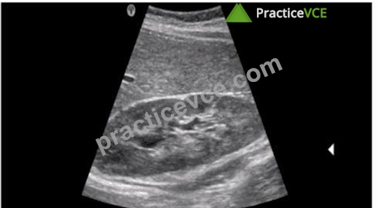
- A. Increase sector width
- B. Decrease depth
- C. Increase dynamic range
- D. Lower focal zone
Answer: C
Explanation:
Dynamic range in ultrasound imaging refers to the range of signal amplitudes that the system can display.
Increasing the dynamic range allows the ultrasound system to display a broader range of echo amplitudes, which enhances the contrast resolution and helps to visualize subtle differences in tissue texture and borders of anatomical structures. When the dynamic range is increased, more shades of gray are used, making the image appear softer and less contrasty, which is beneficial for delineating the borders of anatomical structures more clearly.
American Registry for Diagnostic Medical Sonography (ARDMS). Sonography Principles and Instrumentation (SPI) Examination Review Guide.
NEW QUESTION # 94
Which adjustment will improve visualization of blood flow if color extends outside the boundary of the vessel walls?
- A. Increasing depth
- B. Decreasing wall filter
- C. Decreasing depth
- D. Increasing wall filter
Answer: D
Explanation:
Comprehensive and Detailed Explanation From Exact Extract:
When color "bleeds" outside vessel walls, it often results from motion artifacts or noise from slow tissue motion being displayed as flow. Increasing the wall filter eliminates low-frequency Doppler shifts caused by motion, improving color accuracy within vessel boundaries.
According to sonography instrumentation reference:
"Raising the wall filter eliminates low-velocity signals from vessel wall motion, reducing the appearance of color outside the vessel lumen." Therefore, the correct answer is C: Increasing wall filter.
-
NEW QUESTION # 95
Which adjustment can maintain the same frame rate when the depth is increased?
- A. Increase number of focal zones
- B. Decrease persistence
- C. Decrease image width
- D. Increase frequency
Answer: C
Explanation:
When the depth of imaging is increased, the time it takes for the ultrasound pulses to travel to and from the deeper structures also increases, which can reduce the frame rate. To maintain the same frame rate, one effective adjustment is to decrease the image width. Narrowing the image width reduces the number of scan lines required to create each frame, allowing the system to maintain a higher frame rate despite the increased depth.
ARDMS Sonography Principles and Instrumentation guidelines
Kremkau, F. W. (2015). Diagnostic Ultrasound: Principles and Instruments.
NEW QUESTION # 96
Which factor influences color flow imaging frame rate?
- A. Variance map selection
- B. Line density
- C. Filter selection
- D. Dynamic range
Answer: B
Explanation:
The frame rate in color flow imaging is influenced by several factors, one of the most significant being line density. Line density refers to the number of ultrasound lines used to create an image. Increasing line density improves spatial resolution but requires more time to acquire each frame, thereby reducing the frame rate.
Other factors such as filter selection, dynamic range, and variance map selection can affect the quality of the color flow image, but they do not have as direct an impact on frame rate as line density does.
References:ARDMS Sonography Principles and Instrumentation, Chapter on Color Doppler Imaging.
NEW QUESTION # 97
......
The software is designed for use on a Windows computer. This software helps hopefuls improve their performance on subsequent attempts by recording and analyzing Sonography Principles and Instrumentation (SPI) exam results. Like the actual ARDMS SPI certification exam, Sonography Principles and Instrumentation (SPI) practice exam software has a certain number of questions and allocated time to answer. Any questions or concerns can be directed to the PracticeVCE support team, who are available 24/7. However, the Sonography Principles and Instrumentation (SPI) exam questions software product license must be validated before use.
SPI Materials: https://www.practicevce.com/ARDMS/SPI-practice-exam-dumps.html
- Accurate SPI Exam Questions: Sonography Principles and Instrumentation supply you high-effective Training Brain Dumps - www.vceengine.com ???? Search for { SPI } on ✔ www.vceengine.com ️✔️ immediately to obtain a free download ????SPI Test Dumps
- Free PDF 2026 ARDMS SPI: Marvelous Authentic Sonography Principles and Instrumentation Exam Hub ???? Immediately open ➡ www.pdfvce.com ️⬅️ and search for ⮆ SPI ⮄ to obtain a free download ????Brain SPI Exam
- Latest SPI Exam Simulator ???? Reliable SPI Test Sample ???? SPI Valid Exam Labs ???? Go to website ➡ www.torrentvce.com ️⬅️ open and search for “ SPI ” to download for free ????SPI Free Dump Download
- Free PDF 2026 ARDMS SPI: Marvelous Authentic Sonography Principles and Instrumentation Exam Hub ???? Open ➡ www.pdfvce.com ️⬅️ enter ➥ SPI ???? and obtain a free download ????SPI Reliable Test Prep
- Exam SPI Bible ???? SPI Free Dump Download ???? SPI New Practice Materials ???? Open 《 www.examcollectionpass.com 》 enter 《 SPI 》 and obtain a free download ????SPI Test Preparation
- Excellent ARDMS Authentic Exam Hub – 100% Pass-Rate SPI Materials ???? Search for ▛ SPI ▟ and obtain a free download on ▛ www.pdfvce.com ▟ ????SPI Valid Exam Labs
- Exam SPI Bible ???? Latest SPI Exam Simulator ???? Latest SPI Exam Simulator ???? Open website 《 www.examdiscuss.com 》 and search for ☀ SPI ️☀️ for free download ????Valid SPI Test Dumps
- Free PDF 2026 ARDMS SPI: Marvelous Authentic Sonography Principles and Instrumentation Exam Hub ???? Simply search for “ SPI ” for free download on ☀ www.pdfvce.com ️☀️ ♥SPI New Practice Materials
- SPI Reliable Test Notes ???? SPI Reliable Real Exam ???? Latest SPI Exam Simulator ☎ Search for ➽ SPI ???? and download it for free on ➠ www.pass4test.com ???? website ????Brain SPI Exam
- SPI Test Preparation ???? SPI Reliable Test Prep ❔ SPI New Practice Materials ???? Search for ➠ SPI ???? and easily obtain a free download on ➠ www.pdfvce.com ???? ????SPI Exam Sample
- SPI Pass Leader Dumps ???? SPI Test Dumps ???? Reliable SPI Test Sample ???? Go to website 「 www.troytecdumps.com 」 open and search for ⏩ SPI ⏪ to download for free ????Latest SPI Exam Simulator
- bookmark-vip.com, bookmarkingalpha.com, idavngp439887.estate-blog.com, shaunaqyiq750334.wannawiki.com, push2bookmark.com, orlandoxvil675682.thenerdsblog.com, elijaheoai956406.webbuzzfeed.com, allyourbookmarks.com, www.stes.tyc.edu.tw, nelsonqvwo442093.glifeblog.com, Disposable vapes
P.S. Free & New SPI dumps are available on Google Drive shared by PracticeVCE: https://drive.google.com/open?id=1V_UCURtI2-G4muVwdwMGYXL_h-sw8WnG
Report this wiki page